 English
 Español de México
 Português
 Français
1. Urolithin A The Cellular Target:
DYRK1A is over expressed in DS. This gene is both developmental and functional. This means the disruption present in fetal development continues during the entire lifetime. Heretofore, we have had few options with regard to inhibition of DYRK1a, EGCG and if possible to find it, Harmine. Both can result in liver damage. We chose EGCG but have had to dose it well below the toxicity threshold. It has been helpful but not optimal.
This article includes rational for its use, citations and safety information. Dosages are not yet available for recommendation.
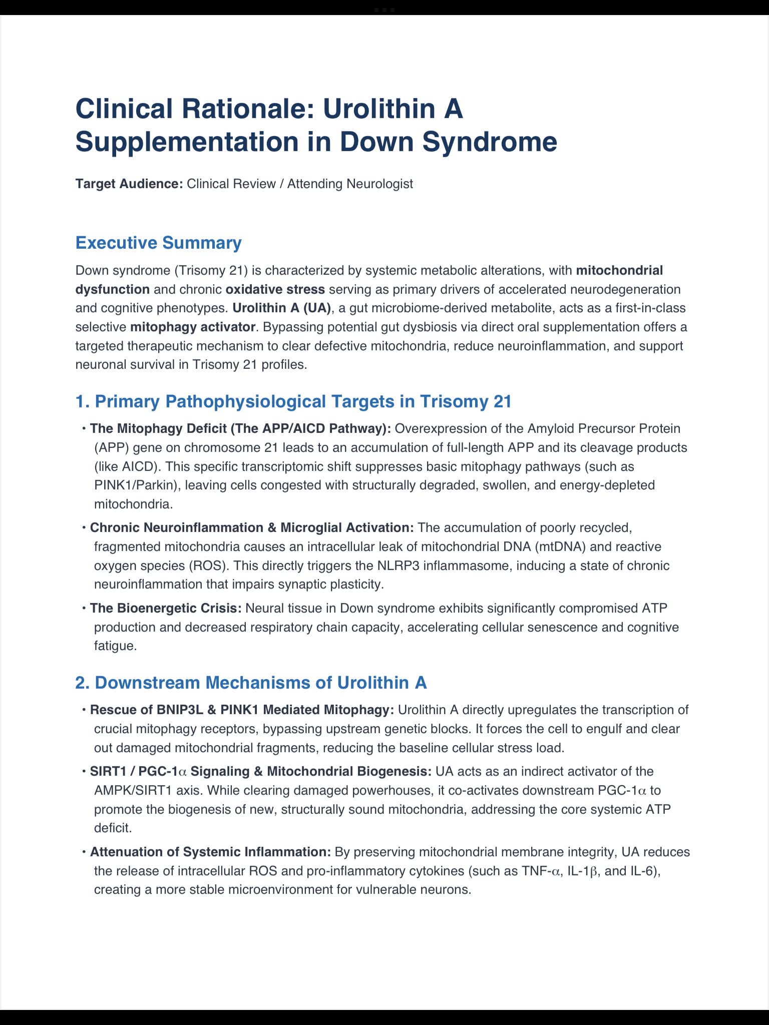
- 1. Overcoming the APP-Driven Mitophagy DeficitThe Down Syndrome Deficit: Due to the triplication of chromosome 21, individuals with Down syndrome have a lifelong overexpression of the Amyloid Precursor Protein (APP) gene. The intracellular accumulation of APP cleavage products (specifically AICD) actively suppresses the cell’s natural PINK1/Parkin and BNIP3L (Nix) mitophagy pathways.The Consequence: Neurons and systemic tissues become congested with swollen, fragmented, and severely compromised mitochondria. This leads to a profound failure in ATP production, severe oxidative stress, and accelerated cellular senescence/cognitive decline.
2. Urolithin A’s Role:
Urolithin A directly activates and rescues downstream BNIP3L-mediated mitophagy. By bypassing the genetic upstream block caused by APP overexpression, UA signals the cell to successfully clear out the toxic mitochondrial fragments.
3. Synergistic Neuroprotective PathwaysAMPK & PGC-1α Activation:
UA stimulates the AMPK/SIRT1 signaling loop. This triggers the downstream expression of PGC-1α, the master driver of mitochondrial biogenesis. This dual action ensures that while old, damaged powerhouses are cleared out, the cell is actively stimulated to synthesize new, highly efficient mitochondria. Calming Microglial “Oxinflammation”: Leakage of mitochondrial DNA (mtDNA) from damaged mitochondria activates the NLRP3 inflammasome in the brain’s immune cells. By stabilizing mitochondrial membranes, UA lowers the chronic pro-inflammatory cytokines (such as TNF-α and IL-6) that hamper synaptic plasticity in Down syndrome profiles.
[ Tau/Microtubules ] [ Cell Cycle/DREAM ] [ Inflammation/Neurogenesis ]
• De-phosphorylates Tau • Blocks Cyclin D1/p27 • Suppresses NF-κB/NLRP3
• Rescues MAP2 • Restores Progenitor Pool • Downregulates IL-6 & TNF-α
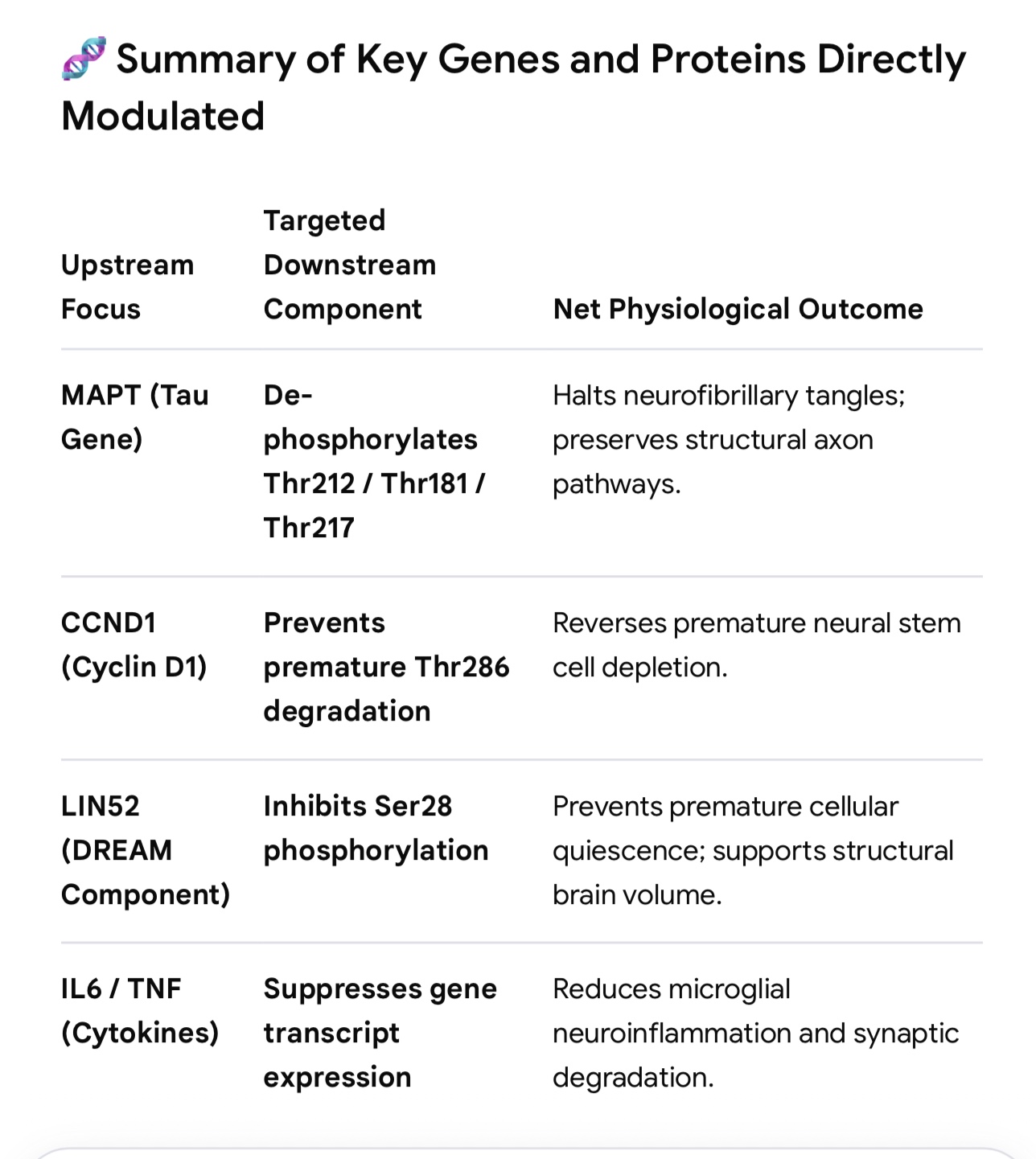
1. The Tau Hyper-phosphorylation & Microtubule Stability PathwayThe Deficit: Excess DYRK1A directly hyper-phosphorylates Tau protein at pathological sites (including Thr181, Thr212, Thr217, and Ser199/202). This causes Tau to detach from microtubules, prompting the formation of neurofibrillary tangles and catastrophic structural collapse of neuronal axons.UA Benefit: By blocking the DYRK1A catalytic pocket, UA triggers immediate tau de-phosphorylation across all major pathological residues. This stabilizes microtubule polymerization, maintains structural cell networks, and preserves axonal transport in vulnerable cortical and hippocampal neurons.
2. The Cell Cycle Quiescence & Neurogenesis Pathway (Cyclin D1 / p27Kip1)The Deficit: In early development and ongoing neurogenesis, DYRK1A over-activation acts as a premature cell-cycle brake. It phosphorylates Cyclin D1 (triggering its destruction) and stabilizes p27Kip1, which forces neural stem cells to exit the cell cycle too early. Furthermore, it hyper-phosphorylates LIN52, driving premature assembly of the DREAM complex and pushing neural precursor pools into permanent dormancy. This underpins the structural brain volume loss and cognitive deficits typical of DS.
UA Benefit: Inhibiting DYRK1A with UA halts the premature degradation of Cyclin D1 and prevents the aberrant assembly of the DREAM complex. This allows neural progenitor cells to remain in the cell cycle long enough to proliferate, expanding the neural stem cell pool and supporting healthier plasticity.
3. The Chronic Synaptic Inflammatory Cascade (NF-κB / NLRP3 / Aβ)
The Deficit: Overexpressed DYRK1A primes microglia and astrocytes into a hyper-reactive state. When exposed to internal structural stressors or early Amyloid-Beta ( ) fragments, over-activated DYRK1A drives the excessive production of destructive inflammatory signals.
UA Benefit: Preclinical profiling shows that UA-mediated inhibition of DYRK1A suppresses -induced microglial activation. This downregulates the transcription of core pro-inflammatory genes, specifically IL-6 and TNF-α, neutralizing the chronic “oxinflammation” that disrupts long-term potentiation (LTP).
4. The Calcineurin-NFAT Transcriptional Axis
The Deficit: DYRK1A works in tandem with RCAN1 (another triplicated gene on chromosome 21) to hyper-phosphorylate NFAT (Nuclear Factor of Activated T-cells) transcription factors. This action pushes NFAT completely out of the nucleus, shutting down genes required for healthy synapse formation and cardiac development.
UA Benefit: Lowering DYRK1A kinase activity allows baseline calcineurin function to safely de-phosphorylate NFAT. This permits its return to the nucleus to resume normal transcription of vital neurodevelopmental genes.
Clinical Advantage Over Traditional Inhibitors
Historically, physicians evaluating DYRK1A targets have relied on compounds like EGCG (from green tea extract). However, EGCG has low bioavailability and faces substantial challenges crossing the blood-brain barrier.
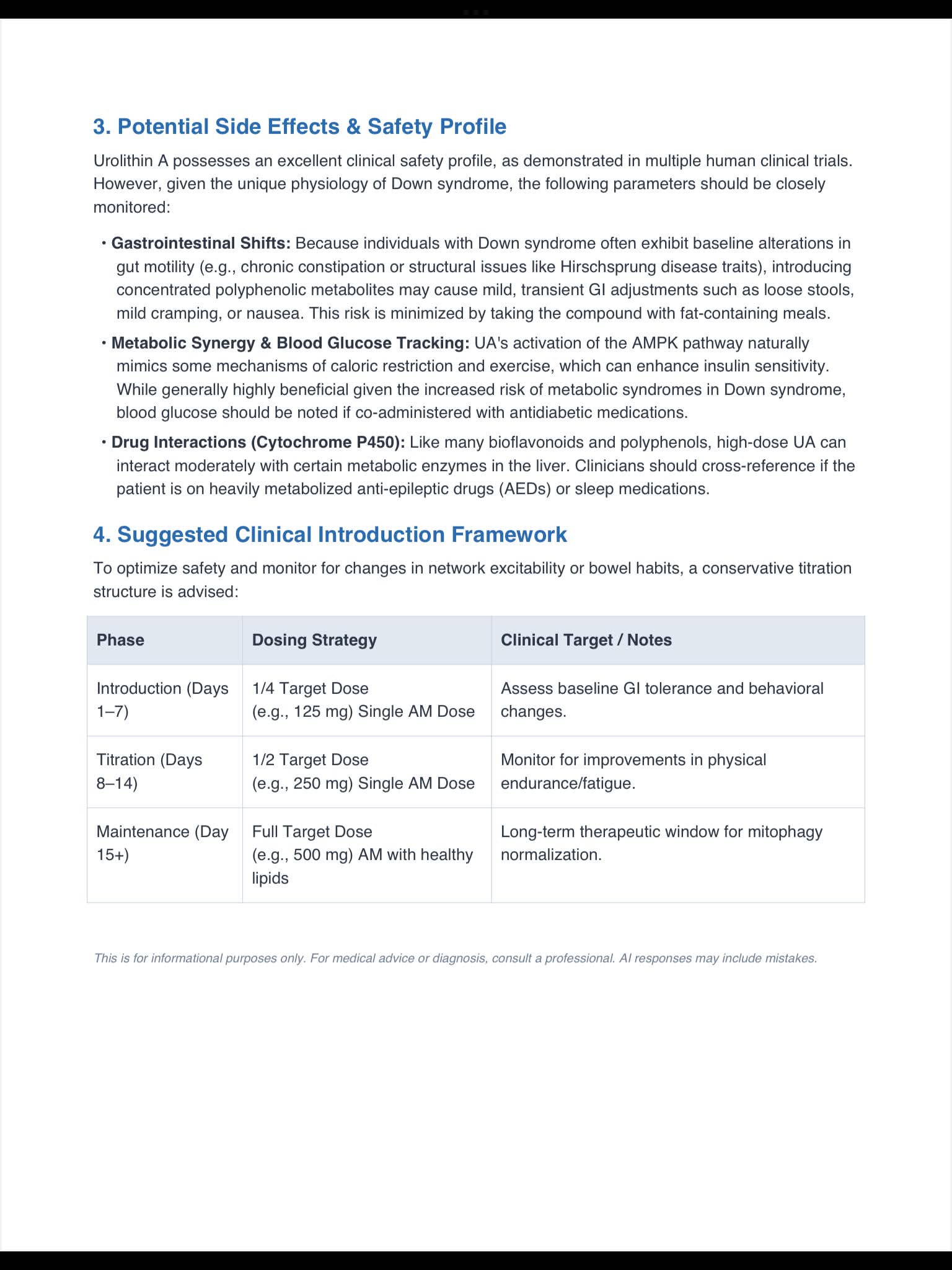
Urolithin A offers a distinct pharmacological profile: it readily crosses the blood-brain barrier, features a stable plasma half-life of 17–22 hours, and targets the cellular engine by concurrently stimulating mitochondrial cleanup (mitophagy). This dual action addresses both the direct genetic over-expression of DYRK1A and the systemic energy failure seen in Down syndrome.
Screenshot
Citations
https://pmc.ncbi.nlm.nih.gov/articles/PMC10281726/
https://pmc.ncbi.nlm.nih.gov/articles/PMC11303307/
https://www.eurekalert.org/news-releases/1121353
https://www.sciencedirect.com/topics/biochemistry-genetics-and-molecular-biology/dyrk1a
https://www.frontiersin.org/journals/molecular-neuroscience/articles/10.3389/fnmol.2024.1391564/full